Tato stránka není v současné době k dispozici ve vašem jazyce. Automatizovaný překlad můžete zobrazit pomocí nástroje Google Translate. Neodpovídáme za poskytování této služby a výsledky překladu jsme nekontrolovali.
Potřebujete-li další pomoc, kontaktujte nás.
Geology
High spectral resolution for complicated structures.
Study varied geological samples
Raman spectroscopy is a great tool for geologists. You can:
- identify minerals and their polymorphic forms
- identify and differentiate other organic and inorganic materials
- determine particle size and distribution in thin sections and polished blocks
- image and identify inclusions within transparent materials
- study samples in different environments, such as at high pressure
- characterise petrochemical samples
-
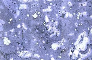
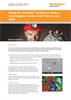
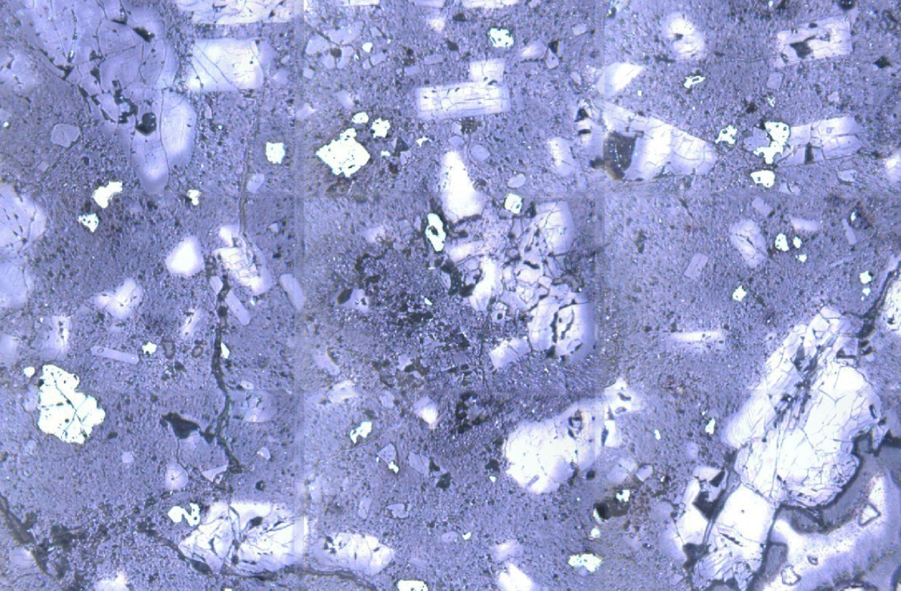
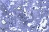
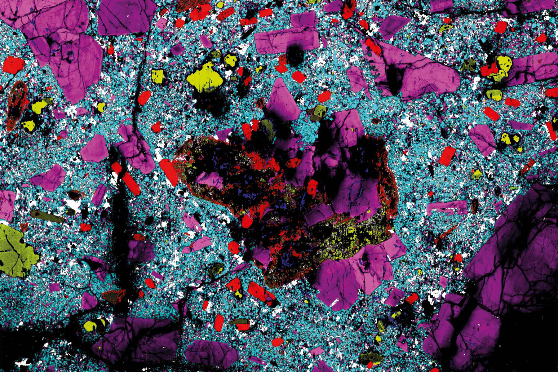
White light image of volcanic rock
-
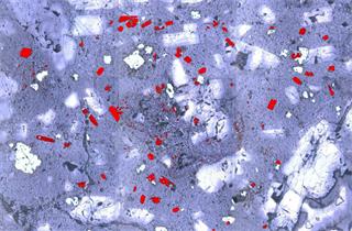
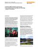
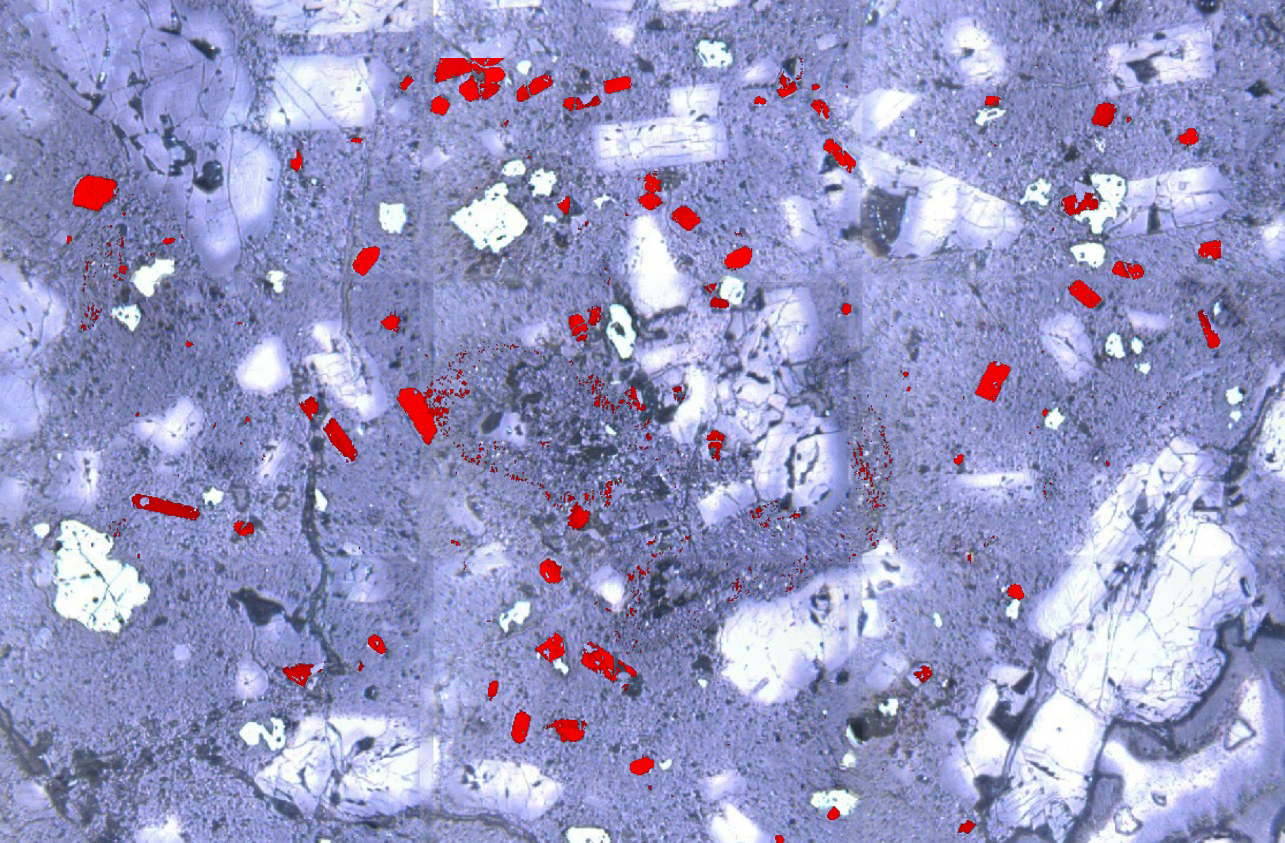
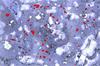
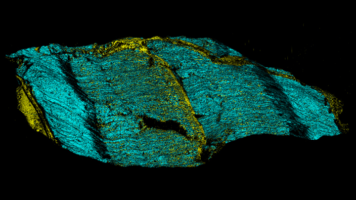
Raman overlay image of volcanic rock
Analyse uneven or rough specimens
See all levels of structure
Geology samples have structures ranging in size from below a micrometre to many millimetres across. Renishaw's inVia confocal Raman microscope can generate images over all these scales.
- High spatial resolution – inVia is highly confocal and can resolve sub-micrometre scale features. You can use an extensive range of high quality microscope objectives—including immersion and cover slip corrected—to reveal small features.
- Large area mapping – Renishaw's HSES motorised stage enables you to analyse large samples, hundreds of millimetres across. Optional open framed free-space microscopes enable you to go larger still.
- High definition images – WiRE software can manage Raman images consisting of over 50 million spectra. You can have high spatial resolution and large area coverage. Capture all the information you need in one image.
Results you can trust
Most minerals are very robust, but some can be transformed by high laser power densities. For example, iron compounds can be oxidised. StreamLine™ uses a laser line, instead of a laser spot. This reduces the laser power density and enables you to analyse sensitive materials without modifying or damaging them.
Easy material identification
You can easily identify materials with Renishaw's Inorganic Materials and Minerals spectral database. The high spectral resolution of the inVia confocal Raman microscope enables you to differentiate material with very similar Raman spectra, such as polymorphs.
Raman+SEM
Add Raman analysis to a scanning electron microscope with Renishaw's inLux™ SEM Raman interface. This instrument is particularly useful for geologists as it combines high spatial resolution SEM images and EDX elemental analysis with the detailed chemical information from Raman spectroscopy.
We're here when you need us
To find out more about this application area, or an application that isn't covered here, contact our applications team.
Contact our applications team
Downloads: earth sciences (geology)
-
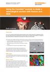 Application note: Using the Correlate™ module to study a mineralogical section with Raman and SEM
An application note detailing the use of the Correlate™ module of the WiRE™ software to combine a Raman and SEM image of the same section of a mineralogical sample and in so doing creating more powerful data, by combining the chemical and physical information of the two techniques.
-
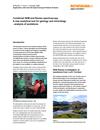 Application note: SCA Geological applications note - sandstone
The SEM-SCA is used to investigate a sandstone sample from the Loch Torridon area of NW Scotland, identifying the component minerals and their phases.